
Corneal Flap Dislocation
LASIK Complications & Risks The first step in the LASIK procedure is creating a corneal flap This is usually performed with a surgical tool called a microkeratome or a laser called IntraLaseA ring is placed on the surface of the cornea and is held in place with suction.

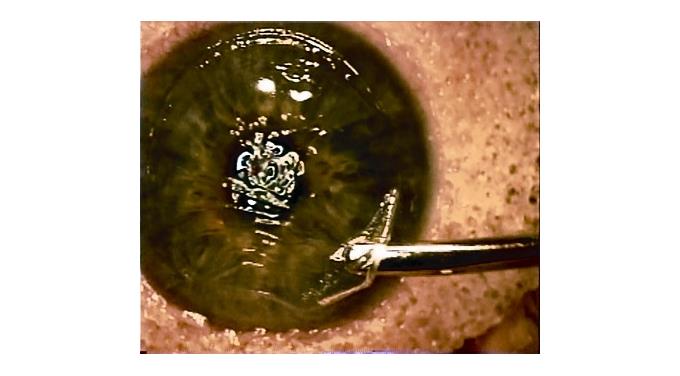
Corneal flap dislocation. Flap dislocation is extremely rare with LASIK even with very powerful injuries including car accidents, sports injuries, and even air big inflation However, the absence of a flap with SMILE makes this an impossibility that may make some people more comfortable with this procedure. The healed flap is stable and unlikely to dislodge with eye rubbing Significant eye trauma could potentially dislocate the flap, however, such instances are rare Flap creation making use of the femtosecond laser provides for even greater stability and reduced chances of flap movement or dislocation. With each dislocation the flap and be prone to develope wrinkling it striae which can reduce quality of vision Placement of sutures is appropriate after the second dislocation but I woukd be concerned about any underlying corneal issues that might be in play with your eye.
LASIK Postoperative complications • Tear instability • Wrinkling, distortion or dislocation of the flap • Subepithelial haze • Persistent epithelial defects • Epithelial ingrowth under the flap • Diffuse lamellar keratitis • Bacterial keratitis • Corneal ectasia. With each dislocation the flap and be prone to develope wrinkling it striae which can reduce quality of vision Placement of sutures is appropriate after the second dislocation but I woukd be concerned about any underlying corneal issues that might be in play with your eye. Despite changes in surgical technique that reduce risk, retina specialists still encounter corneal defects with vitrectomy and intravitreal injectionsRetinal Physician editor in chief Peter K Kaiser, MD, leads a discussion with retina specialists Carl D Regillo, MD, FACS, and Thomas A Albini, MD, and cornea specialists Marguerite McDonald, MD, FACS, and Francis S Mah, MD, to try to reach.
This case illustrates that dislocation of corneal flap may happen following blunt trauma even when the corneal epithelial cells are supposed to have regenerated 19 days after LASIK AB A 48yearold female received uneventful laser in situ keratomileusis (LASIK) for myopia in both eyes. Late LASIK flap dislocation cases have been reported up to 14 years after surgery 2 Although a rare event, with enough force a LASIK corneal flap may be completely avulsed, and sometimes loss Searching the MEDLINE literature database through PubMed interface we found that only 12 cases have been reported in the English literature ( Table 1 ) 3 , 4 , 5 , 6 , 7 , 8 , 9. Despite changes in surgical technique that reduce risk, retina specialists still encounter corneal defects with vitrectomy and intravitreal injectionsRetinal Physician editor in chief Peter K Kaiser, MD, leads a discussion with retina specialists Carl D Regillo, MD, FACS, and Thomas A Albini, MD, and cornea specialists Marguerite McDonald, MD, FACS, and Francis S Mah, MD, to try to reach.
Corneal flap tears and dislocations In 05, Yeh et al published a case using fibrin glue and mechanical debridement to treat traumatic LASIK flap dislocation and epithelial ingrowth The patient had bilateral LASIK 21 months prior to flap dislocation After treatment,. The flap does not dislocate as the very edge is not cut through (making it a flap) The surgeon smooths the corneal flap down after the correction is completed It then adheres and heals in a short amount of time if the corneal flap dislocates after the procedure it can be felt by the patient and is quite uncomfortable. These observations clearly illustrate that corneal integrity is never fully restored after creation of a LASIK flap The basis for this inherent weakness has recently been reported in a rabbit model of LASIK in which irregular corneal stromal regeneration was observed at the wound margin 5 Flap dislocation as a potential late complication.
Seven years after uneventful laser in situ keratomileusis (LASIK), a 48yearold woman presented one week after being hit with an iron cord with blurry vision, pain, and irritation The injury resulted in traumatic flap dislocation, epithelial ingrowth, and macrostriae Following epithelial removal, the flap was refloated and repositioned Nine interrupted sutures were used to secure the flap. Many cases have been reported regarding late onset flap dislocation and related problems 13 To our knowledge, however, this is the first case report of interfacecaptured foreign bodies after a mild corneal scratch without any sign of flap displacement The foreign bodies were associated with Staphylococcal keratitis. Microkeratomerelated flap complications Flap Buttonhole caused by buckling of the cornea during flap creation, occurs predominantly in steep corneas Other risk factors include loss of suction, defective blade, abnormal advancement of blade Management Do not perform laser ablation, recut the flap and ablate a minimum of 3 months later Free Cap.
Flap wound never heals completely, said Dr Schocket, who’s experienced a dislocation twice In fact, studies have shown that the strength of the healed wound margin is on average only 28% of the normal cornea1 This can lead to flap dislocation during retina surgery “I’ve learned to specifically ask patients. CLEAR is a laser corneal surgery treatment that corrects myopia and astigmatism The expert surgeons at CODET Vision Institute offer outpatient CLEAR surgery for quick and painless vision correction. A LASIK flap wound never heals completely, said Dr Schocket, who’s experienced a dislocation twice In fact, studies have shown that the strength of the healed wound margin is on average only 28% of the normal cornea 1 This can lead to flap dislocation during retina surgery “I’ve learned to specifically ask patients multiple times whether they’ve had LASIK, not just eye surgery,” she said, “because many don’t think of it as a surgical procedure” (See “Dislocation of a.
However, this may result in significant irregular astigmatism 7. Corneal flap tears and dislocations In 05, Yeh et al published a case using fibrin glue and mechanical debridement to treat traumatic LASIK flap dislocation and epithelial ingrowth The patient had bilateral LASIK 21 months prior to flap dislocation After treatment,. Two interventional case reports of patients with lateonset LASIK corneal flap dislocation after ocular trauma occurring at 7 and 26 months after surgery, respectively The flaps were lifted.
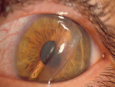
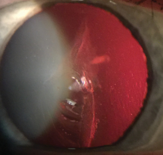
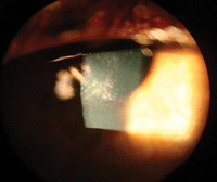
A, B, C color photograph of a dislocated laser in situ keratomileusis flap 24 days after surgery secondary to a “finger flick” to the right eye (patient 2) Note the severe folds across the visual axis (B) as well as the coverage of the exposed stroma by corneal epithelium (C) within 10 hours of the injury. The flap dislocation in our case was caused by a twig 10 years after the LASIK procedure Epithelial ingrowth into the interface between the flap and stromal bed is a complication of LASIK flap dislocation Epithelial ingrowth with direct communication and supply from peripheral regenerating epithelium could lead to progressive ingrowth under. The precise cause of flap dislocation is unclear, but wrinkling or dislodgment may result from poor adherence of the corneal flap to the stromal bed because of overhydration of the stromal bed or.
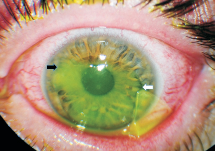
Rationale Traumatic flap dislocation might occur anytime after laser in situ keratomileusis (LASIK), but it is rarely concomitantly complicated with epithelial ingrowth, infectious keratitis, and diffuse lamellar keratitis altogether Here we report a case of traumatic LASIK flap inversion with epithelial ingrowth, Propionibacterium acnes infection, and diffuse lamellar keratitis. Examination, he had a 180degree traumatic corneal flap dislocation with an inverted tear in the temporal cornea (Panel A, fluorescein dye) After repositioning of the flap, a disposable contact lens, used as a therapeutic bandage, was placed on the cornea. Traumatic corneal flap dislocation one to six years after LASIK in nine eyes with favorable outcome Print J Refract Surg 06 Nov;22(9)49 Landau D, Levy J, Solomon A, Lifshitz T, Orucov F, Strassman E, FruchtPery J.
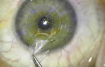
Late flap dislocations are always associated with ocular trauma, though most ocular trauma does not cause a dislocation, thus accounting for their significantly reduced frequency compared to early dislocations The LASIK flap actually consists of two wounds (1) the planar interface between the flap and the underlying corneal stroma and (2) the. Before repositioning the dislocated flap, the ophthalmologist will evaluate the stromal surfaces of the flap and the corneal stroma should be scraped to remove any debris and epithelial cells In the event of a lost flap, the epithelium is simply allowed to heal;. Specifically, the corneal flap created during LASIK can become dislocated following minor physical trauma, such as a finger or object striking the eye If the flap becomes dislocated, the patient would require further medical or surgical intervention to correct the dislocation and protect the patients vision1.
Corneal flap displacement within 1 to 2 days following LASIK is a wellrecognized complication1–3However, stability of the flap remains largely unknown as flap dehiscence has been reported as late. Dislocated intraocular lens (IOL) is a rare, yet serious complication whereby the intraocular lens moves out of its normal position in the eye IOL dislocation has been reported at a rate of 02% to 3% 14 It may occur as a result an early or late complication of cataract surgery, prior vitreoretinal surgery, trauma, or an inherent pathological process or connective tissue disorders. Delayed diagnosis and management of flap dislocation after corneal trauma may potentially increase the risk of epithelial ingrowth, recalcitrant flap striae and visual impairment The presence of lateonset epithelial ingrowth in patients with previous LASIK mandates careful examination for occult flap displacement.
The precise cause of flap dislocation is unclear, but wrinkling or dislodgment may result from poor adherence of the corneal flap to the stromal bed because of overhydration of the stromal bed or the flap, eye rubbing, excessive blinking, and eye squeezing from pain, photophobia, or other discomfort in the postoperative period. 2 Flap Related Complication If the corneal flap is not made correctly with mechanical microkeratome, its adherence to the corneal bed will not be good in which it might get wrinkled, folded or dislocated Flap dislocation in this case can occur spontaneously or after minor trauma or eye rubbing Treatment of it should be surgical. As described earlier, the flapstromal interface is vulnerable given the limited healing that occurs While flap dislocation or penetrating cornea trauma introduces an entrance into this potential space for foreign body debris, another possible mechanism involves sharp ocular trauma that may leave foreign body debris in the LASIK interface.
Here’s a general rule to live by with regard to corneal flap dislocation If you’re not sure whether or not your corneal flap has moved, then your corneal flap has not moved In the very unlikely event of corneal flap dislocation, patients will feel immediate pain and should seek emergency treatment. Corneal transplant LASIK permanently thins and thereby weakens the cornea This can lead to the need for a cornea transplant, despite a LASIK surgery being deemed “successful” Corneal flap dislocation The corneal flap is altered during every LASIK surgery However, in some cases, this eventually leads to a dislocated corneal flap. Dislocated intraocular lens (IOL) is a rare, yet serious complication whereby the intraocular lens moves out of its normal position in the eye IOL dislocation has been reported at a rate of 02% to 3% 14 It may occur as a result an early or late complication of cataract surgery, prior vitreoretinal surgery, trauma, or an inherent pathological process or connective tissue disorders.
During LASIK, a small flap is created in the topmost layer of the corneal, which is known as the epithelium By moving this top layer of the cornea back, a LASIK surgeon can then reshape and recontour the corneal surface and improve the passage of light through the eye Once the corneal reshaping is commplete, the flap is set back down. Examination, he had a 180degree traumatic corneal flap dislocation with an inverted tear in the temporal cornea (Panel A, fluorescein dye) After repositioning of the flap, a disposable contact lens, used as a therapeutic bandage, was placed on the cornea. The Lasik flap dislocation is a separation between layers of collagen within the cornea and it does adhere back to the underlying collagen after few weeks of surgery especially the flaps made by using femtosecond laser In simple terms, it actually adheres quite well if the surgery is performed with the use of modern technology.
Late flap dislocations are always associated with ocular trauma, though most ocular trauma does not cause a dislocation, thus accounting for their significantly reduced frequency compared to early dislocations The LASIK flap actually consists of two wounds (1) the planar interface between the flap and the underlying corneal stroma and (2) the. Here’s a general rule to live by with regard to corneal flap dislocation If you’re not sure whether or not your corneal flap has moved, then your corneal flap has not moved In the very unlikely event of corneal flap dislocation, patients will feel immediate pain and should seek emergency treatment. All the cases with corneal flap dislocation had received flap replacement (Table 5), while only 556% of cases in Xiao et al study had undergone the same treatment Lack of ophthalmic doctors skilled in refractive surgery could be one possible.
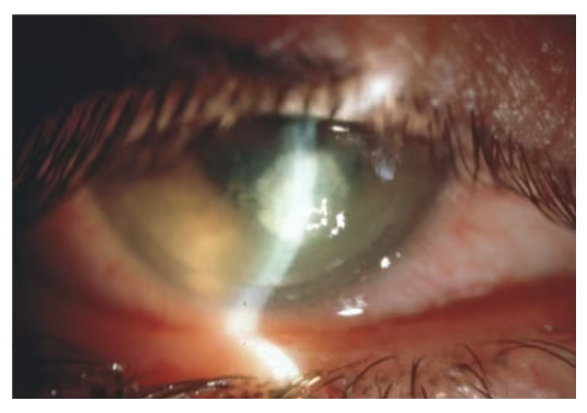
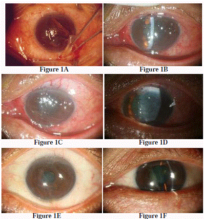
I have an article published in this month's Journal of Refractive Surgery titled Traumatic Dislocation of LASIK Flaps 4 and 9 Years After Surgery With the help of UMDNJ ophthalmology resident, H Jane Kim, we published two cases of traumatic flap dislocation following LASIK One of the cases occurred 9 years following LASIK, making it the longest reported case medical literature. The pictures of the corneal flap dislocation in our patient showed a larger upper portion of the corneal flap compared to the lower portion Furthermore, there was a part of the upper portion cornea folding between the stromal bed and cornea itself Besides, the patient had multiple lacerations over upper eyelids. Specifically, the corneal flap created during LASIK can become dislocated following minor physical trauma, such as a finger or object striking the eye If the flap becomes dislocated, the patient would require further medical or surgical intervention to correct the dislocation and protect the patients vision1.
These observations clearly illustrate that corneal integrity is never fully restored after creation of a LASIK flap The basis for this inherent weakness has recently been reported in a rabbit model of LASIK in which irregular corneal stromal regeneration was observed at the wound margin 5 Flap dislocation as a potential late complication. The precise cause of flap dislocation is unclear, but wrinkling or dislodgment may result from poor adherence of the corneal flap to the stromal bed because of overhydration of the stromal bed or. Corneal flap displacement within 1 to 2 days following LASIK is a wellrecognized complication 1–3 However, stability of the flap remains largely unknown as flap dehiscence has been reported as.
The microkeratome or IntraLase then creates a flap leaving a small hinge to keep the flap partially attached to the rest of the cornea Research suggests the incidence of LASIK complications with flaps is about 02 percent (0002) of all LASIK surgeries (Study of Corneal flap complications from Codet Aris Vision Institute). A superiorhinged corneal flap was created using the Moria M2 microkeratome (Moria SA, Antony, France) and the surgery was uneventful Ten years later, partial flap dislocation was diagnosed after mild trauma This case suggests that flap dislocations can occur during recreational activities up to 10 years after surgery. This technique reduces the risk of corneal edema because it requires no irrigation under the flap;.
The Lasik flap dislocation is a separation between layers of collagen within the cornea and it does adhere back to the underlying collagen after few weeks of surgery especially the flaps made by using femtosecond laser. Corneal transplant LASIK permanently thins and thereby weakens the cornea This can lead to the need for a cornea transplant, despite a LASIK surgery being deemed “successful” Corneal flap dislocation The corneal flap is altered during every LASIK surgery However, in some cases, this eventually leads to a dislocated corneal flap. Purpose To report the management and outcome of late onset traumatic dislocation of LASIK flaps Methods This retrospective, interventional case series presents three patients with late onset LASIK flap dislocation following mechanical trauma 1 to 7 years postoperatively Results In all cases, the flap was surgically repositioned.
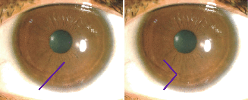
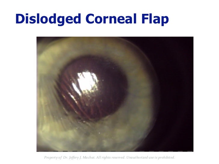
There have been cases of striae reported without corneal flap dislocation following trauma years after the initial surgery It is important to consider the formation of striae when examining LASIK patients with ocular trauma even in the absence of flap slippage, subluxation, or dislocation Fluorescein dye can be helpful to detect flap folds. Flap Melt The flap begins to disintegrate and may require surgical intervention Flap Dislocation This is a condition in which the corneal flap becomes dislodged and no longer properly aligns with the corneal bed This is treated with surgical intervention Flap Wrinkles A major or minor flap dislocation produces flap wrinkles This is. A flap dislocation would cause notable pain, discomfort, excessive watering in the eye, and/or blurred vision The vast majority of flap dislocation incidents happen in the first few days after the LASIK procedure as a result of not wearing eye protection and being hit or bumped in the eye.
Two interventional case reports of patients with lateonset LASIK corneal flap dislocation after ocular trauma occurring at 7 and 26 months after surgery, respectively The flaps were lifted. Purpose To describe postoperative laserassisted in situ keratomileusis (LASIK) flap dislocation occurred after trauma Methods Ultrabiomicroscopy (UBM) is used to obtain a highresolution imaging of the cornea Results The UBM results are presented and compared with histologic and confoscan findings Conclusions The technique is useful and easy to perform, offering more opportunities to. The flap dislocation in our case was caused by a twig 10 years after the LASIK procedure Epithelial ingrowth into the interface between the flap and stromal bed is a complication of LASIK flap dislocation Epithelial ingrowth with direct communication and supply from peripheral regenerating epithelium could lead to progressive ingrowth under.
The precise cause of flap dislocation is unclear, but wrinkling or dislodgment may result from poor adherence of the corneal flap to the stromal bed because of overhydration of the stromal bed or the flap, eye rubbing, excessive blinking, and eye squeezing from pain, photophobia, or other discomfort in the postoperative period. All the cases with corneal flap dislocation had received flap replacement (Table 5), while only 556% of cases in Xiao et al study had undergone the same treatment Lack of ophthalmic doctors skilled in refractive surgery could be one possible. Seven years after uneventful laser in situ keratomileusis (LASIK), a 48yearold woman presented one week after being hit with an iron cord with blurry vision, pain, and irritation The injury resulted in traumatic flap dislocation, epithelial ingrowth, and macrostriae Following epithelial removal, the flap was refloated and repositioned Nine interrupted sutures were used to secure the flap.
Edema increases the risk of the flap's not fitting well into the stromal bed and of epithelial defects, both of which are associated with striae, flap dislocation, and DLK.
Lasik And Flap Complications Salt Lake City Ut Risks

Corneal Flap Dehiscence After Screwdriver Trauma Nejm
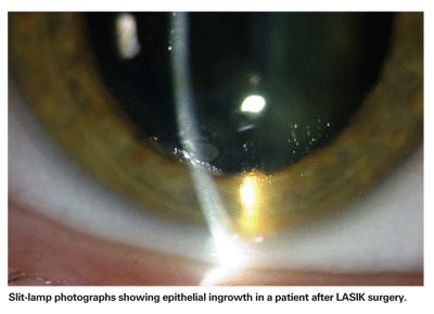
Treating Epithelial Ingrowth After Lasik
Corneal Flap Dislocation のギャラリー
/PRK%20Step%203.png)
Refractive Surgery Lasik

Small Incision Lenticule Extraction Smile It S What S New In Laser Vision Correction Harvard Health Blog Harvard Health Publishing

Treatment Of Traumatic Lasik Flap Dislocation And Epithelial Ingrowth With Fibrin Glue American Journal Of Ophthalmology

The Lasik Vision Institute Flap Dislocation After Lasik
Q Tbn And9gcsuqogvtezopkkbaabxhlouhhh4fbcqyb9wrltk7rogroagnb8g Usqp Cau

Eyeworld Management Of Rare Post Lasik Complications

Lasik Complications And Their Management Ento Key
.jpg)
Lasik Flap Dislocation The Flap Never Heals

Z Ruo9rcshewlm

Late Post Traumatic Flap Dislocation And Macrostriae After Laser In Situ Keratomileusis Sinha R Shekhar H Tinwala S Gangar A Titiyal Js Oman J Ophthalmol
Lasik Laser Eye Surgery
Lasik Prk Eye Surgery Fredericksburg King George Stafford Va

Lasik Springfield Il Lasik Eye Surgery Springfield Prairie Eye
Http Link Springer Com Content Pdf 10 1007 2f978 3 540 5 4 Pdf
Intraoperative Flap Complications In Lasik Prevention And Management Of Free Flaps Springerlink

Epithelial Ingrowth After Late Traumatic Femtosecond Laser Assisted Laser In Situ Keratomileusis Flap Dislocation Sciencedirect
.jpg)
Lasik Flap Dislocation The Flap Never Heals
Early Adopter Chronic Sufferer
Http Www Jkslms Or Kr Journal Download Pdf Php Doi 10 252 Ml 16 5 1 47
Late Traumatic Flap Dislocation Seven Years After Femtosecond Laser Assisted In Situ Keratomileusis

Circumferential Epithelial Defect At Flap Margins In Patient With Adenoviral Conjunctivitis And Previous Lasik Eye

Slit Lamp Photograph Of The Injured Eye Five Days After Flap Download Scientific Diagram
Flap Complications From Femtosecond Laser Assisted In Situ Keratomileusis Touchophthalmology
2

Lasik Interface Primary Complications
Early Lt 3 Months And Late Gt 3 Months Complications Of Lasik Springerlink

Top 10 Reasons Why You Should Not Get Lasik Eye Surgery Omg Top Tens List

Lasik Interface Primary Complications

Eye Sight Fluctuations 2 Weeks After Lasik Any Suggestions Photos

Ophthalmology Management Refractive Surgery With A Smile
Refractive Surgery Complication Management By Dr Jeff Machat

Lasik For Correction Of Myopia Treatment Management Preoperative Details Intraoperative Details Postoperative Details

Pdf Management Of A Traumatic Flap Dislocation Seven Years After Lasik
Dislocated Corneal Lamellar Flap Following Blunt Ocular Injury A Case Report

A Step By Step Approach To The Diagnosis And Management Of Sands Of Sahara Syndrome Eye News
Www Ecronicon Com Ecop Pdf Ecop 10 Pdf

Jaypeedigital Ebook Reader

Traumatic Corneal Flap Displacement Five Years After Laser In Situ Keratomileusis Semantic Scholar

Patients A Guide To Lasik Prk And Smile Tfos Tear Film Ocular Surface Society

Full Text Traumatic Corneal Flap Displacement After Laser In Situ Keratomileusis Imcrj
Full Text Traumatic Corneal Flap Displacement After Laser In Situ Keratomileusis Imcrj

Management Of A Traumatic Flap Dislocation Seven Years After Lasik

Which Is Better Lasik Or Prk Schulze Vision Surgery Center
Q Tbn And9gctxxqxjjpvvi4xlgrxpldxjizal5f2ir T5evlptk1oe3wovlvu Usqp Cau
Late Traumatic Flap Dislocation Seven Years After Femtosecond Laser Assisted In Situ Keratomileusis
Crstoday Vision Loss After Post Lasik Trauma
.jpg)
Lasik Flap Dislocation The Flap Never Heals
Q Tbn And9gcrkrxzaxwr Uozxmhbkdijxoacnwepafe6yzmjt6 2d2u6mq Cn Usqp Cau

Crstoday How Should I Manage The 10 Year Post Lasik Patient Who Is Now Unhappy With His Or Her Vision
Www Touchophthalmology Com Wp Content Uploads Sites 16 19 04 Us Ophth 12 1 P21 27 1 Pdf

Lasik Vs Prk What The Lasik Doctor Doesn T Tell You Is This Your Homework

Sandra Lora Cremers Md Facs What To Ask Before You Have Lasik Or Prk In 17 Lasik Risks And Complications

Late Traumatic Flap Dislocation Seven Years After Femtosecond Laser Assisted In Situ Keratomileusis Abstract Europe Pmc

Femtosecond Laser Eases Lasik Fears

Late Traumatic Lasik Flap Loss During Contact Sport Sciencedirect

Recovery Time After Laser Eye Surgery What To Expect Dougherty Laser
Late Traumatic Flap Dislocation Seven Years After Femtosecond Laser Assisted In Situ Keratomileusis

Pdf Traumatic Corneal Flap Dislocation Four Years After Lasik Semantic Scholar
2
Www Healio Com Ophthalmology Cornea External Disease Journals Osli 08 5 39 3 7b68b97a92 53d3 4a13 87b9 Fff463aa9990 7d Successful Delayed Surgical Revision Of A Dislocated Lasik Flap Pdf
Late Onset Dehiscence Of Lasik Flap Poster Number P 190 Authors Hyunjin Jane Kim Cary M Silverman Category Keratorefractive Authors Have No Financial Ppt Download
Flap Complications Of Lasik Ask The Eye Doctor Webeyeclinic
:max_bytes(150000):strip_icc()/GettyImages-895653602HowLongDoesLasikSurgeryLast-c666fa0115154513bbe5b6d42cd019f0.jpg)
Permanent Or Temporary How Long Does Lasik Last

Traumatic Flap Dislocation 10 Years After Lasik Case Report And Literature Review Sciencedirect

Management Of A Traumatic Flap Dislocation Seven Years After Lasik
Refractive Surgery Goes Intrastromal
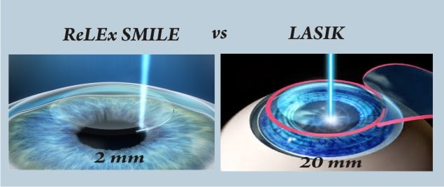
Difference Between Relex Smile Lasik Laser Procedure

Full Text Traumatic Corneal Flap Displacement After Laser In Situ Keratomileusis Imcrj

Eyeworld Smile Compared To Lasik

Therapeutic Flap Amputation For Atypical Lasik Flap And Interface Abnormalities
Lasik Jonesblog

Services Mahajan Eye Centre

Lasik Flap Stability After Severe Ocular Injury Sciencedirect

Escrs Journal Of Cataract And Refractive Surgery

The Resurgence Of Post Lasik Epithelial Ingrowth William Nc Nixon Hk Pudukadan D Bhat L Kerala J Ophthalmol

A Clear Look At The Lasik Flap
/flap_edge_dye(425).jpg)
Lasik Flap Dislocation The Flap Never Heals
/ectasia_flap_margin(425).jpg)
Lasik Flap Dislocation The Flap Never Heals
.jpg)
Lasik Flap Dislocation The Flap Never Heals
Lvc No Longer Out Of Bounds For Athletes
Www Tandfonline Com Doi Pdf 10 1586 Eop 11 8

Ophthalmology Management How To Manage Femto Lasik Complications

Lasik Laser Eye Surgery Safety Recovery Time Complications

Pdf Management Of A Traumatic Flap Dislocation Seven Years After Lasik
/detached-lasik-flap(640).jpg)
Lasik Flap Dislocation The Flap Never Heals

Slit Lamp Photograph Of The Injured Eye Five Days After Flap Download Scientific Diagram

Pdf Traumatic Corneal Flap Dislocation Four Years After Lasik Semantic Scholar
Lasik Complications Corneal Ectasia Subconjunctival Hemorrhage Halos Around Lights

Late Post Traumatic Flap Dislocation And Macrostriae After Laser In Situ Keratomileusis Sinha R Shekhar H Tinwala S Gangar A Titiyal Js Oman J Ophthalmol

Femtolasik The Bladeless Eye Surgery Magrabi Hospitals

Eyeworld Management Of Rare Post Lasik Complications
Laser Eye Surgery Primer Lasik Vs Prk Is This Your Homework
Www Ecronicon Com Ecop Pdf Ecop 10 Pdf

Figure 4 From Satisfactory Clinical Outcome Following Delayed Repositioning Of A Traumatic Post Lasik Flap With Dislocation And Shrinkage Managed By Irrigation Stretching And Debridement Semantic Scholar

Late Traumatic Dislocation Of Laser In Situ Keratomileusis Corneal Flaps Ophthalmology

Smile Vs Lasik
Q Tbn And9gcs8bcqllwzmk8fhrg6b9qibae06rdxy9 Epmnf8k9kuvgjxvokf Usqp Cau

What Makes Femto Lasik The Safest Laser Eye Surgery

Lasik Complications And Their Management Ento Key

Troubleshooting In Lasik Eye News

Epithelial Ingrowth Following Laser In Situ Keratomileusis Lasik Prevalence Risk Factors Management And Visual Outcomes Bmj Open Ophthalmology


