Flap Corneal Arrugado
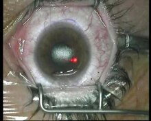
LASIK or Lasik (laserassisted in situ keratomileusis), commonly referred to as laser eye surgery or laser vision correction, is a type of refractive surgery for the correction of myopia, hyperopia, and astigmatism LASIK surgery is performed by an ophthalmologist who uses a laser or microkeratome to reshape the eye's cornea in order to improve visual acuity.

Flap corneal arrugado. A third eyelid flap is not recommended as it provides no nutritive or structural support to the eye, may cause irritation, and prevents close monitoring of the ulcer Corneal ulcer patients should wear an appropriately fitted, hard plastic ecollar at all times to reduce the likelihood of selftrauma as well as the chance of secondary infection. A su lado, sobre una silla, había un raído sombrero de copa y un gabán marrón descolorido, con el arrugado cuello de terciopelo En resumidas cuentas, y por mucho que yo lo mirase, nada de notable distinguí en aquel hombre, fuera de su pelo rojo vivísimo y la expresión de disgusto y de pesar extremados que se leía en sus facciones. When performing all laser LASIK, our physicians use a gentle laser to create the corneal flap This technology, also used to facilitate laser cataract surgery, allows them to better customize the corneal flap for each patientAs the name states, no blades are used in this kind of laser eye surgery.
Al día siguiente de la cirugía el paciente tendrá que volver con el fin de revisarle y examinar su visión. El 17 de Mayo me opere ambos ojos (LASIK) para corregir miopía y astigmatismo, al tercer día veía mejor con el ojo derecho que el izquierdo, a los 10 días asistí al primer control y me dijeron que se me había arrugado el flap corneal del OI, así que. Flap y realizar una intervención exitosa Otras veces el flap corneal queda ligeramente arrugado o el tratamiento con el láser no está totalmente centrado con la pupila, requiriendo una reintervención para solucionarlo con éxito La cirugía refractiva para USARLA COMO UN RETOQUE está indicada en pacientes que tengan más de 18 años y una.
Does it heal only at the edges or the entire flap under surface heals and attaches itself back to the treated corneal base How strong is the healed bond?. De la misma. Conclusions Conjunctival flap cover surgery is an underused technique Its primary indication is refractory corneal ulcer and corneal perforation, and its second indication is aesthetic with poor visual potential coexisting ocular surface diseases It represents an interesting alternative to more mutilating surgeries.
The corneal flap will selfadhere itself naturally in just a few minutes This flap serves as a protective covering for the area of the eye that has been operated on, and thus facilitates rapid healing The corneal flap begins healing immediately after the LASIK procedure In fact, with the use of a LASIK flap, the corneal tissue can be as much. THE TECHNIQUE of using a conjunctival flap for the treatment of chronic corneal ulceration was described by Gunderson 1 in the late 1950s and became a standard surgical procedure There are some drawbacks to this procedure, however 1,2 During the past years, progress in microsurgical technique, aided by the microscope and fine surgical tools, has enabled us to use a selective pedunculated. See what Alia Edrees (aliaedrees) has discovered on , the world's biggest collection of ideas.
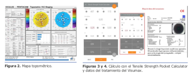
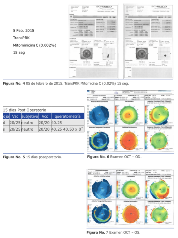
En esta sesión de ALACCSAR deseamos presentar unos casos complicados de cirugía refractiva o “el peor” caso de cirugía refractiva Se trata de cirujanos con mucha experiencia que describen complicaciones que sucedieron en años recientes y que probablemente son muy infrecuentes y otras obedecen a la aplicación de nuevas tecnologías. Measurement of the residual stromal bed beneath a LASIK flap on ASOCT The central corneal thickness is estimated to be 525µm, and the residual stromal bed is measured to be 321µm centrally and µm toward the peripheral cornea At the University of Iowa, a patient is deemed not a candidate for LASIK or enhancement if the final. Purpose To assess the efficacy and safety of transepithelial photorefractive keratectomy (TPRK) without mitomycin C as treatment for femtosecond laser in situ keratomileusis (FSLASIK) corneal flap complications Methods Eight patients with corneal flap complications that occurred after FSLASIK (five with eccentric flaps, two with buttonhole flaps, and one with a thick flap) were included.
Diathermy of large feeding vessels and corneal laser photocoagulation has been among the surgical modalities to treat pannus Limbal grafting may be required in eyes with severe chemical injuries and limbal epithelium loss. Corneal hysteresis (CH), corneal resistance factor (CRF), central corneal thickness (CCT), Goldmanncorrelated intraocular pressure (IOPg), and cornealcompensated intraocular pressure (IOPcc. Sus ojos estarán muy sensibles y no hay que perturbar la cicatrización del flap corneal en caso de haber sido operado de FemtoLasik.
Conclusions Conjunctival flap cover surgery is an underused technique Its primary indication is refractory corneal ulcer and corneal perforation, and its second indication is aesthetic with poor visual potential coexisting ocular surface diseases It represents an interesting alternative to more mutilating surgeries. Translator Translate texts with the world's best machine translation technology, developed by the creators of Linguee Linguee Look up words and phrases in comprehensive, reliable bilingual dictionaries and search through billions of online translations. Corneal Endothelium – An extremely thin layer, this is the innermost layer of the cornea These cells are necessary in order to keep the cornea transparent If any injury breaks through to a deeper layer of the cornea, the healing process takes longer and may result in the following symptoms blurred vision, pain, redness, sensitivity to.
Aim To characterise temporal changes in corneal wound repair at the LASIK flap margin Methods 18 rabbits received monocular LASIK and were evaluated during 6 months using slit lamp and in vivo confocal microscopy In three corneas, the exposed stroma was stained with DTAF At various time points, corneas were processed for histology and stained for nuclei, factin, EDA fibronectin, α. Corneal Imaging An Introduction Miles F Greenwald, BS, Brittni A Scruggs, MD, PhD, Jesse M Vislisel, MD, Mark A Greiner, MD October 19, 16 Introduction Imaging techniques for assessing the structure and function of the cornea and anterior segment are crucial for diagnosing and treating a wide variety of ocular diseases. The next frame illustrates the corneal flap being lifted and folded back over on its hinge The next illustration shows the eye at an angle with the corneal flap laid open In a smaller frame.
A bridge flap may be created with a narrow band of conjunctiva that runs across the corneal lesion allowing a greater view of the anterior chamber Edges of the flap overlying the cornea are sutured with 100 nylon and those overlying episclera with 80 polyglactin 910 (eg Vicryl) Nonabsorbable suture knots must be buried. Aug 24, 18 Explore maison dárt's board "Royal Blue Color" on See more ideas about royal blue color, designers guild, blue color. Guía de campo de las babosas marinas del Pacífico Este Tropical 05 Terrence Gosliner.
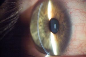
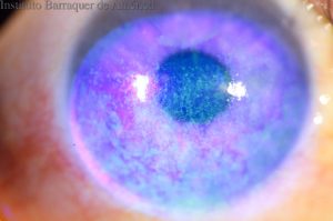
Purpose To describe the morphological characteristics of microfolds that appear at the corneal flap after LASIK, as seen under confocal microscopy Methods Twentyone eyes that had undergone LASIK were examined, all within 3 weeks to 1 month after surgery A central scan of the total corneal thickness was obtained by using confocal microscopy in vivo. Flap striae have multiple etiologies Causes include the improper placement of the flap onto the corneal bed, as well as a deep photoablation resulting in tenting of the flap in the bed and folds induced by the normal loss of tissue Moreover, excessive flap manipulation may produce an irregular flap and cause corneal striae. Corneal abrasions are one of the most common forms of eye injury In some cases, they are caused by the direct impact of a sharp object, such as a pencil, staple, nail or sewing pin They also can be caused by small, airborne particles, such as dust, sand or flying debris from soldering, woodworking or weed trimming.
Simulation of corneal flaps The corneal flap refers to a thin flap of corneal tissue that is cut during refractive surgery In our study, the diameter of the corneal flap was 81mm, and the thickness of the corneal flap was presumed to be 90, 1, 150, 180, 210, and 240μm, respectively (Fig 2) Corneal shape change and stress distribution. ¿Es normal ver borroso después de un desarrugue del flap corneal?. Dec 31, 13 Explore Nathan M's board "Steampunk Mens Clothing" on See more ideas about steampunk, steampunk clothing, steampunk costume.
El 17 de Mayo me opere ambos ojos (LASIK) para corregir miopía y astigmatismo, al tercer día veía mejor con el ojo derecho que el izquierdo, a los 10 días asistí al primer control y me dijeron que se me había arrugado el flap corneal del OI, así que el 31 de Mayo me programaron desarrugue donde me dijeron que tuvieron que raspar un poco de cornea y me colocaron un lente de contacto por. ARRUGADO One method for preparing the binder for production, where the tobacco is folded in creases ASC An Internet cigar forum, where participants discuss a wide variety of cigar related information One can be a ‘lurker’, that is, passive, reading the postings, or be an active participant The subjects are provocative, at times. A corneal inlay is a tiny device, smaller than the width of an eraser tip During a 1015 minute surgery, a laser is used to cut a small pocket or flap in the middle of the nondominant eye’s cornea The inlay is then inserted into or under this pocket or flap, depending on which inlay is used.
Flap y realizar una intervención exitosa Otras veces el flap corneal queda ligeramente arrugado o el tratamiento con el láser no está totalmente centrado con la pupila, requiriendo una reintervención para solucionarlo con éxito La cirugía refractiva para USARLA COMO UN RETOQUE está indicada en pacientes que tengan más de 18 años y una. An icon used to represent a menu that can be toggled by interacting with this icon. The desired corneal flap thickness for both groups was 160 μm In the 4mmHg group, corneal flap thickness was 1600 ± 62 μm (range, 1523–1674 μm);.
A su lado, sobre una silla, había un raído sombrero de copa y un gabán marrón descolorido, con el arrugado cuello de terciopelo En resumidas cuentas, y por mucho que yo lo mirase, nada de notable distinguí en aquel hombre, fuera de su pelo rojo vivísimo y la expresión de disgusto y de pesar extremados que se leía en sus facciones. Introduction Corneal refractive laser surgery is considered to be safe, predictable and efficient in treatment of myopia with or without astigmatism (Wen et al 17)The most commonly performed corneal refractive procedure worldwide is laser‐assisted keratomileusis (LASIK) developed by Pallikaris (Pallikaris et al 1990, 1991)Here, a corneal hinged flap is cut with a microkeratome or. Soy conciente que el superficie tiene que ser bastante duro, pero es como no hubiera nada de relleno dentro Se desliza mucho, incluso si esta encima de una alfombra, así al aprender gatear mi peque acaba con el gimnasio arrugado debajo de el y no va a ningún lado.
What Are Corneal Inlays?. What Are Corneal Inlays?. Free cap is a rare intraoperative complication of LASIK (laser in situ keratomileusis) Ideally in LASIK, a hinged corneal flap is created that allows eximer laser to be applied on the exposed stromal bed If the hinge of the corneal flap detaches, the flap becomes a free flap/cap The occurrence of this complication is most commonly associated with flat corneas, which predisposes to small.
LASIK Flap Recovery After Procedure The corneal flap will begin the healing process immediately following the surgery and will be significantly healed in one to three days following the procedure During this time the outer surface of the cornea (also known as the epithelium) will seal the edges of the newly created corneal flap. However, significant trauma can occur which requires ophthalmic examination and treatment. 1 Introduction The laser in situ keratomileusis (LASIK) procedure for correcting refractive errors consists of the creation of a corneal flap, ablation of the stroma by excimer laser, and reposition of the flapCreating a corneal flap poses risks to the integrity of the corneal surface The lowgrade wound healing between the corneal flap and the stromal bed maintains corneal transparency.
Ese mismo día deberá ser capaz de ducharse sin que le entre agua, jabón o cualquier otro tipo de producto en los ojos. After reshaping the exposed cornea with an excimer laser, the surgeon must reposition the corneal flap back to its original position over the stromal bed, usually using sponges and forceps. Corneal Trauma Introduction The anterior segment of the eye is constantly exposed to minor trauma throughout the normal day The precorneal tear film, corneal epithelium, and conjunctival epithelium provide protection against this continued mild trauma;.
Creating the Corneal Flap To create the corneal LASIK flap, the LASIK surgeon uses a handheld device or a laser to cut the top layers of corneal tissue at a predetermined depth One edge of the flap is left uncut, forming a hinge Using the hinge, the surgeon folds back the flap to access the underlying corneal tissue After the surgeon has. Surgical management of traumatic corneal flap epithelial ingrowth and macro striae. Corneal opacities are eye problems that can lead to scarring or clouding of the cornea, which decreases vision The cornea is the clear, domeshaped area that covers the front of the eyeLight.
Corneal flap Smoothly polished biconvex surfaces are designed for entering the interface, lifting and repositing the flap Can be used to squeeze excess fluid from the interface K Maddox LASIK Spatula– combines a flat spatula for elevating the flap and protecting the hinge with a cylindrical spatula for repositing and smoothing the flap. The corneal flap begins healing immediately after the LASIK procedure In fact, with the use of a LASIK flap, the corneal tissue can be as much as 90% healed within 24 hours During the first day or two after surgery, the outer surface of the cornea, known as the epithelium, seals the edges of the corneal flap Over the next few weeks, natural. A bridge flap may be created with a narrow band of conjunctiva that runs across the corneal lesion allowing a greater view of the anterior chamber Edges of the flap overlying the cornea are sutured with 100 nylon and those overlying episclera with 80 polyglactin 910 (eg Vicryl) Nonabsorbable suture knots must be buried.
I am 26 and have worn glasses for distance vision for the last 15 years I had been considering laser eye surgery for a very long time and finally went through with it last Thursday (Nov 10, 11) The surgery itself was as painless as everyone had said it would be I was given Valium and numbing eye drops The surgeon. LASIK Flap Recovery After Procedure The corneal flap will begin the healing process immediately following the surgery and will be significantly healed in one to three days following the procedure During this time the outer surface of the cornea (also known as the epithelium) will seal the edges of the newly created corneal flap. The microkeratome or IntraLase then creates a flap leaving a small hinge to keep the flap partially attached to the rest of the cornea Research suggests the incidence of LASIK complications with flaps is about 02 percent (0002) of all LASIK surgeries (Study of Corneal flap complications from Codet Aris Vision Institute).
The corneal flap could obviously cause a deformation of the anterior corneal surface For example, the displacement of the corneal vertex was 15 μm more than that without the corneal flap when the corneal flap was 1 μm thick The issue of cutting depth in refractive surgery was not considered in our study and would be explored in the future. Gundersen Conjunctival Flap Contributors Jesse M Vislisel, MD and Mark A Greiner, MD September 24, 15 The Gundersen conjunctival flap procedure involves the transposition of a thin flap of conjunctiva to cover the cornea for the relief of painful ocular surface disorders or to provide metabolic support for corneal healing. During LASIK, a small flap is created in the topmost layer of the corneal, which is known as the epithelium By moving this top layer of the cornea back, a LASIK surgeon can then reshape and recontour the corneal surface and improve the passage of light through the eye Once the corneal reshaping is commplete, the flap is set back down.
Aug 24, 18 Explore maison dárt's board "Royal Blue Color" on See more ideas about royal blue color, designers guild, blue color. The flap involves making an incision in the circular strip of the outer corneal tissue, then separating it from the underlying stroma However, one segment of the tissue remains attached by not completing the circle – thus, a small portion of the cornea acts like a hinge Once the tissue is cut to size, the ‘flap’ is complete. In case of any potential accident or rubbing the eyes will not dislodge the flap right?.
After completing the conjunctival flap dissection, create the partialthickness corneal–scleral trabeculectomy flap, which is the guarded part of this procedure If a limbalbased technique was used, gently reflect the conjunctival flap over the cornea and gently hold it with smooth forceps or a WeckCel sponge. How does corneal flap heals after Lasik?. Introduction Corneal refractive laser surgery is considered to be safe, predictable and efficient in treatment of myopia with or without astigmatism (Wen et al 17)The most commonly performed corneal refractive procedure worldwide is laser‐assisted keratomileusis (LASIK) developed by Pallikaris (Pallikaris et al 1990, 1991)Here, a corneal hinged flap is cut with a microkeratome or.
In the 600mmHg group, the flap thickness was 1246 ± 1324 μm, (range, 14–2662 μm) as shown in Table 2 The differences were not statistically significant. After completing the conjunctival flap dissection, create the partialthickness corneal–scleral trabeculectomy flap, which is the guarded part of this procedure If a limbalbased technique was used, gently reflect the conjunctival flap over the cornea and gently hold it with smooth forceps or a WeckCel sponge. The flap is lifted like a hinged door, targeted tissue is removed from the corneal stroma, again with the microkeratome, and then the flap is replaced Laserassisted in situ Keratomileusis (LASIK) The surgeon uses either a microkeratome or a femtosecond laser to cut a flap of the corneal tissue (usually with a thickness of 100–180 micrometres).
The incidence of a corneal melt following LASIK is difficult to quantify accurately, as reports in published literature are often small series or case reports discussing a single event 1 The melting process often starts at the rim of the flap and is commonly associated with a variety of conditions, such as epithelial ingrowth 1 The migration. A corneal inlay is a tiny device, smaller than the width of an eraser tip During a 1015 minute surgery, a laser is used to cut a small pocket or flap in the middle of the nondominant eye’s cornea The inlay is then inserted into or under this pocket or flap, depending on which inlay is used. Purpose To analyse the accuracy of corneal flap thickness created in laser‐assisted in situ keratomileusis (LASIK) using the Moria Model 2 (M2) single‐use head 90 microkeratome Methods The corneal thickness of 300 (266 myopic and 34 hyperopic) eyes of 150 patients was measured by ultrasonic pachymetry preoperationally and intraoperationally after flap cut.

Pdf Graphene Based 3d Scaffolds In Tissue Engineering Fabrication Applications And Future Scope In Liver Tissue Engineering

Tratamiento De Pliegues Del Lenticulo Tras Cirugia Lasik

Ro 5ta 12 By Creative Latin Media Llc Issuu
Flap Corneal Arrugado のギャラリー
Lasik Wikipedia La Enciclopedia Libre

Prk Lasek Introduccion Y Complicaciones Afectados Cirugia Refractiva

Casos Complicados De Cirugia Refractiva Alaccsa R

Pdf Graphene Based 3d Scaffolds In Tissue Engineering Fabrication Applications And Future Scope In Liver Tissue Engineering

Lasik Flap Wrinkles Corrected Graphic Content Viewer Discretion Advised Youtube

Pdf Graphene Based 3d Scaffolds In Tissue Engineering Fabrication Applications And Future Scope In Liver Tissue Engineering

Lasik Wikipedia La Enciclopedia Libre

10 Actos A Evitar Despues De Una Cirugia Laser Vallmedic Vision

Es Normal Ver Borroso Despues De Un Desarrugue Del Flap Corneal Vista Todoexpertos Com
Lens Absorption Fotos E Imagenes De Stock Alamy

Lens Absorption Fotos E Imagenes De Stock Alamy
Diccionario Siderurgico Ingenieria Naturaleza
Las Complicaciones En La Cirugia Corneal Refractiva Alaccsa R

Lasik Introduccion Y Complicaciones Afectados Cirugia Refractiva

Lasik Introduccion Y Complicaciones Afectados Cirugia Refractiva

Despues De La Cirugia Ocular Con Laser Los Ojos Estaran Danados De Por Vida Afectados Cirugia Refractiva
Mis Lentes De Contacto Se Mueven Y Veo Borroso Vista Todoexpertos Com

Pdf Graphene Based 3d Scaffolds In Tissue Engineering Fabrication Applications And Future Scope In Liver Tissue Engineering

Tratamiento A Largo Plazo De Macroestrias Post Laser In Situ Keratomileusis Mediante Queratectomia Fototerapeutica Transepitelial Caso Clinico

Recuperacion De La Vision Tras Una Cirugia Laser Vallmedicvision

Como Hacer Mascarillas Hidratantes Para La Cara Las Mas Efectivas Hidratante Para La Cara Mascarillas De Aguacate Piel Seca En La Cara
Casos Complicados De Cirugia Refractiva Alaccsa R

10 Actos A Evitar Despues De Una Cirugia Laser Vallmedic Vision

Pdf Graphene Based 3d Scaffolds In Tissue Engineering Fabrication Applications And Future Scope In Liver Tissue Engineering

Despues De La Cirugia Ocular Con Laser Los Ojos Estaran Danados De Por Vida Afectados Cirugia Refractiva
Casos Complicados De Cirugia Refractiva Alaccsa R

Lasik Flap Wrinkles Corrected Graphic Content Viewer Discretion Advised Youtube

Ebook Dic Spanish English Dictionary 19 466 Entries 2 Nature
Las Complicaciones En La Cirugia Corneal Refractiva Alaccsa R

Lasik Flap Wrinkles Corrected Graphic Content Viewer Discretion Advised Youtube

Lasik Wikipedia La Enciclopedia Libre

Lasik Introduccion Y Complicaciones Afectados Cirugia Refractiva

Lasik Flap Wrinkles Corrected Graphic Content Viewer Discretion Advised Youtube

Cirugia Refractiva Postoperatorio Y Dudas Informacion De Opticas
Recuperacion De La Vision Tras Una Cirugia Laser Vallmedicvision

Cirugia Refractiva Postoperatorio Y Dudas Informacion De Opticas

Diccionario Para Ingenieros Ingles Espanol Queroseno Acero

Tratamiento De Pliegues Del Lenticulo Tras Cirugia Lasik
Que Es Una Arruga O Pliegue Macular American Academy Of Ophthalmology

Lasik Flap Wrinkles Corrected Graphic Content Viewer Discretion Advised Youtube

Lasik Wikipedia La Enciclopedia Libre

Pdf A Ch Orti Morphological Sketch Soren Wichmann Academia Edu

Luna Cornea 29 Maravilla By Centro De La Imagen Issuu

Tratamiento De Pliegues Del Lenticulo Tras Cirugia Lasik

Lasik Introduccion Y Complicaciones Afectados Cirugia Refractiva
Diccionario Para Ingenieros Publicacion Naturaleza

Tratamiento A Largo Plazo De Macroestrias Post Laser In Situ Keratomileusis Mediante Queratectomia Fototerapeutica Transepitelial Caso Clinico

The Pathogenesis Of Floppy Eyelid Syndromeinvolvement Of Matrix Metalloproteinases In Elastic Fiber Degradation Request Pdf

Lasik Flap Wrinkles Corrected Graphic Content Viewer Discretion Advised Youtube

Diccionario Siderurgico Ingenieria Naturaleza

Lasik Flap Wrinkles Corrected Graphic Content Viewer Discretion Advised Youtube

Lasik Presente Y Futuro Ablacion A La Medida Con Frente De Onda Pdf Oftalmologia Vision

Lasik Introduccion Y Complicaciones Afectados Cirugia Refractiva

Pdf Graphene Based 3d Scaffolds In Tissue Engineering Fabrication Applications And Future Scope In Liver Tissue Engineering
Recuperacion De La Vision Tras Una Cirugia Laser Vallmedicvision

Tratamiento De Pliegues Del Lenticulo Tras Cirugia Lasik
Lens Absorption Fotos E Imagenes De Stock Alamy
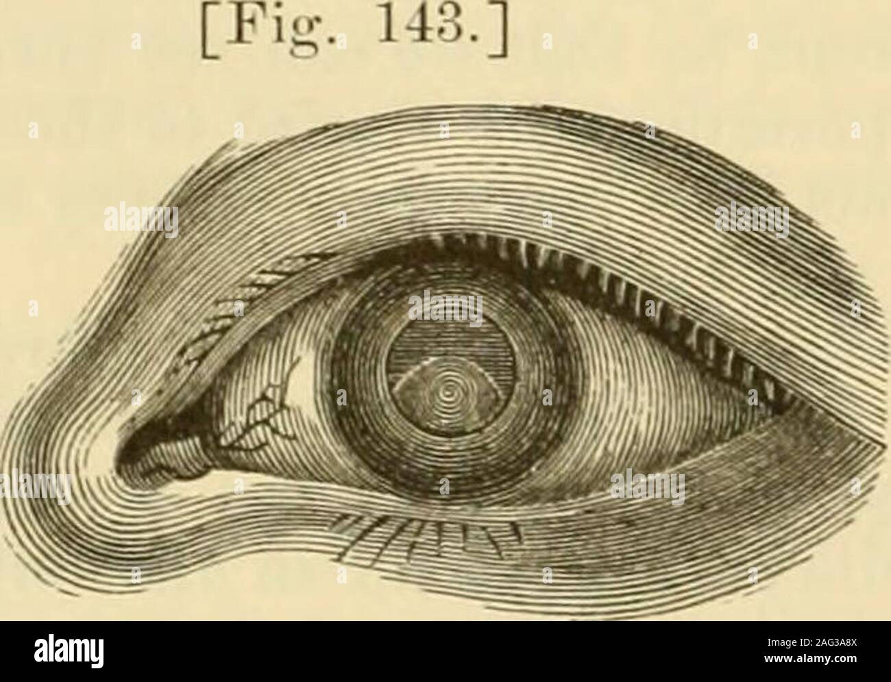
Lens Absorption Fotos E Imagenes De Stock Alamy
Frotarse Los Ojos Es Malo

Diccionario Para Ingenieros

Recuperacion De La Vision Tras Una Cirugia Laser Vallmedicvision

Despues De La Cirugia Ocular Con Laser Los Ojos Estaran Danados De Por Vida Afectados Cirugia Refractiva

Despues De La Cirugia Ocular Con Laser Los Ojos Estaran Danados De Por Vida Afectados Cirugia Refractiva

Tratamiento De Pliegues Del Lenticulo Tras Cirugia Lasik

Lasik Introduccion Y Complicaciones Afectados Cirugia Refractiva


