
Corneal Flap Displacement
The revolutionary IntraLase® system removes the element of even slight human error, resulting in corneal flaps that have perfectly straight edges, with strong hinges and uniform depths Although using the IntraLase® system to create corneal flaps does not eliminate the possibility of flap complications, it reduces them dramatically.
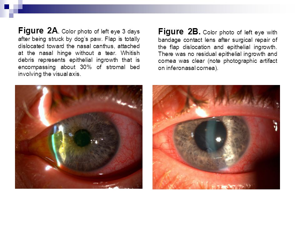
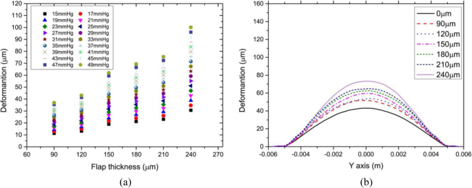
Corneal flap displacement. There have been reports of corneal flap displacement up to 38 months after the procedure After surgery, women are cautioned to not wear eye makeup or use lotions and creams around the eyes for a minimum of two weeks and to discard all previously used makeup to reduce the risk of infection. RESULTS The results showed that the corneal flap could obviously cause the deformation of the anterior corneal surface For example, the displacement of the corneal vertex achieved 15 μm more than that without corneal flap, when the thickness of corneal flap was 1 μm thick. Partial displacement of the corneal flap may result in corneal striae or wrinkles, which blur vision Most striae are treatable, but some patients, such as those who are highly nearsighted, may be resistant to treatment Complete displacement of the corneal flap is often painful and requires urgent replacement.
The creation of a lamellar flap results in a potential plane of weakness in the cornea in which shearing forces can produce flap displacement Recent histological and confocal studies have shown a central hypocellular primitive scar in the interface, allowing easy lifting of the flap in trauma. Laser in situ keratomileusis (LASIK) is a relatively new ophthalmic procedure that represents a combination of previously used techniques in refractive surgery1 It involves using a microkeratome to create a thin corneal flap, followed by excimer laser stromal ablation and repositioning of the flap2 The creation of a corneal flap represents additional risks of intraoperative and postoperative. Corneal Flap Displacement Young Hee Yoon MD Posted 5/08/14 Keywords Laser refractive Surgery, Vision Correction, LASIK.
Displacement of a corneal fl ap after LASIK isa serious complication which has been identified after corneal epithelial debridement duringa scleral buckling procedure and vitrectomy Itis extremely important for the refractive surgeonto detect and treat certain conditionsbefore LASIK, including misdiagnosed posteriorpole diseases such as. All patients had peripheral corneal edema in their recipient bed All 3 patients required an additional surgical procedure for visual rehabilitation Conclusion Flap displacement may occur following LASIK in patients who have undergone PKP for bullous keratopathy. The corneal flap following LASIK is initially held in place by suction created by the pumping function of the endothelium against an intact epithelium Any evidence of epithelial or stromal corneal edema is a sign that the endothelial pump function has been reduced below the natural swelling pressure of the cornea, and the adherence of the flap.
During LASIK, a small flap is created in the topmost layer of the corneal, which is known as the epithelium By moving this top layer of the cornea back, a LASIK surgeon can then reshape and recontour the corneal surface and improve the passage of light through the eye Once the corneal reshaping is commplete, the flap is set back down. Traumatic corneal flap displacement after laser in situ keratomileusis (LASIK) By Tsai TsungHan, KaiLing Peng and ChienJen Lin Cite BibTex;. The corneal flap could obviously cause a deformation of the anterior corneal surface For example, the displacement of the corneal vertex was 15 μm more than that without the corneal flap when the corneal flap was 1 μm thick The issue of cutting depth in refractive surgery was not considered in our study and would be explored in the future.
Publisher Dove Medical Press Ltd Year 17 DOI identifier /imcrjs OAI identifier Provided by MUCC. A late onset of corneal flap displacement was found with upper and lower portion of the flap being folded inside the corneal bed Surgical intervention for debridement with subsequent reposition of corneal flap was performed as soon as possible in the operating room. Displacement of a corneal flap after LASIK is a serious complication which has been identified after corneal epithelial debridement during a scleral buckling procedure and vitrectomy It is extremely important for the refractive surgeon to detect and treat certain conditions before LASIK, including misdiagnosed posterior pole diseases such as.
The flap involves making an incision in the circular strip of the outer corneal tissue, then separating it from the underlying stroma However, one segment of the tissue remains attached by not completing the circle – thus, a small portion of the cornea acts like a hinge Once the tissue is cut to size, the ‘flap’ is complete. Laser in situ keratomileusis (LASIK) is a relatively new ophthalmic procedure that represents a combination of previously used techniques in refractive surgery1 It involves using a microkeratome to create a thin corneal flap, followed by excimer laser stromal ablation and repositioning of the flap2 The creation of a corneal flap represents additional risks of intraoperative and postoperative. Traumatic corneal flap displacement is an uncommon complication followinglaserassisted in situ keratomileusis (LASIK) Most reported traumatic flapdisplacements are partial and can subsequently be repositioned with satisfactoryresults 14 Occasionally,however, significant force may completely avulse the corneal flap, resultingin a lost flap 5,6 Completeloss of the flap may cause significant.
Displacement of a corneal fl ap after LASIK isa serious complication which has been identified after corneal epithelial debridement duringa scleral buckling procedure and vitrectomy Itis extremely important for the refractive surgeonto detect and treat certain conditionsbefore LASIK, including misdiagnosed posteriorpole diseases such as. PURPOSE To describe and compare the displacement of corneal flaps created during laser in situ keratomileusis (LASIK) procedures performed with two different microkeratomes and analyze parameters (for example, pupiltohinge distance, drift during suction) that might affect the flap displacement or be influenced by flap displacement. Partial displacement of the corneal flap may result in corneal striae or wrinkles, which blur vision Most striae are treatable, but some patients, such as those who are highly nearsighted, may be resistant to treatment Complete displacement of the corneal flap is often painful and requires urgent replacement.
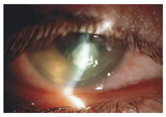
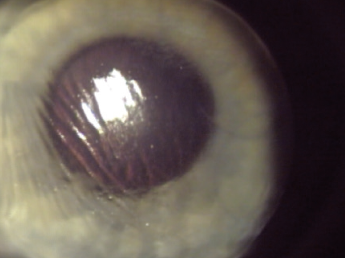
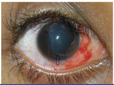
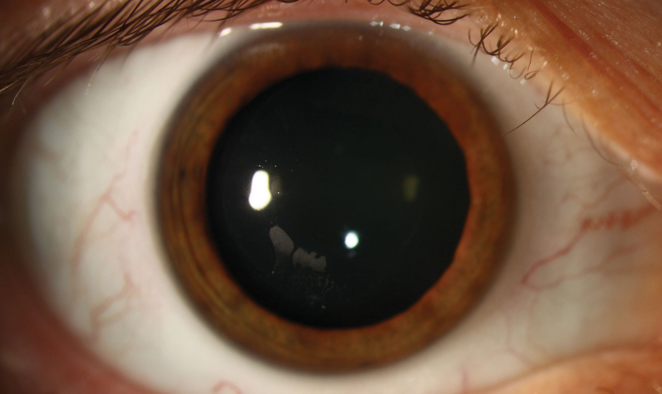
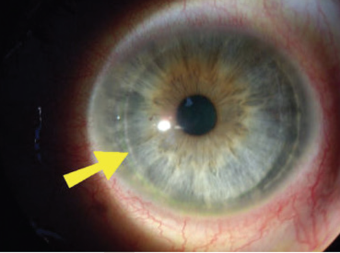
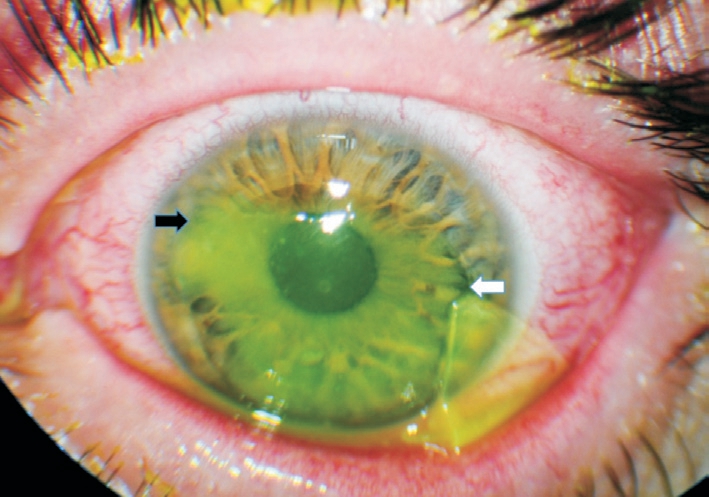
A lateonset corneal flap displacement with superior and inferior portion of flap being folded is shown Bare stroma is exposed and intact nasal hinge is visible under surgical microscope. Flap vs Lens , , and have to do with the intraocular lens not the corneal flap done for LASIK For the corneal flap dislocation I use 998. Displacement of a corneal flap after LASIK is a serious complication which has been identified after corneal epithelial debridement during a scleral buckling procedure and vitrectomy It is extremely important for the refractive surgeon to detect and treat certain conditions before LASIK, including misdiagnosed posterior pole diseases such as.
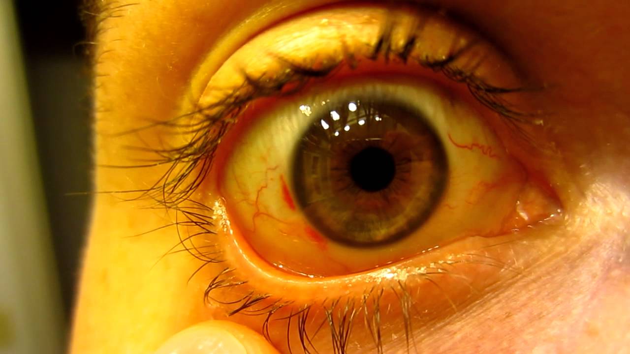
Relationship between the anterior corneal surface displacement along the Yaxis and corneal flap thickness The corneal flap thickness was assumed to be 90, 1, 150, 180, 210, and 240 μ m. Dr Jess Mason presents a case of a patient with a history of LASIK eye surgery who got poked in the eye, causing displacement of her corneal flap The flap was irrigated, gently replaced, and she was started on antibiotic eye drops with close ophthalmology follow up. The first critical step in LASIK is creation of the corneal flap The LASIK flap is paramount to the procedure and allows for quick visual recovery with little pain, 10 but is also a major source of complications There are two main methods for creating corneal flaps Early flap displacement after LASIK.
For example, the displacement of the corneal vertex achieved 15μm more than that without corneal flap, when the thickness of corneal flap was 1μm thick This displacement was enough to cause the change of aberrations in the human eyes In the central part of the cornea, the stress on the anterior corneal surface increased with flap thickness. Traumatic displacement of corneal flaps after LASIK may occur after blunt injury with specific direction of force to the flap margin, especially tangential one According to the previous literature, lateonset traumatic flap displacement may happen at any time after LASIK and be caused by various types of injuries. Corneal Flap Displacement Young Hee Yoon MD Posted 5/08/14 Keywords Laser refractive Surgery, Vision Correction, LASIK.
Creating a corneal flap poses risks to the integrity of the corneal surface The lowgrade wound healing between the corneal flap and the stromal bed maintains corneal transparency, but on the other hand, leads to weak flap adhesion Flap dislocation might occur any time even as long as 14 years after LASIK Here we report a case of traumatic. The corneal flap begins healing immediately after the LASIK procedure In fact, with the use of a LASIK flap, the corneal tissue can be as much as 90% healed within 24 hours During the first day or two after surgery, the outer surface of the cornea, known as the epithelium, seals the edges of the corneal flap Over the next few weeks, natural. Flap displacement – To reshape the corneal tissue, a flap is created over the cornea to access into the cornea and it is repositioned after the surgery But if the flap folds back or gets removed then this condition is known as flap displacement and it can cause serious problem.
Keywords traumatic flap displacement, laser in situ keratomileusis, LASIK, corneal flap, reposition, debridement Introduction Traumatic flap displacement, which is known as one of the complications of LASIK, may occur in the early or late postoperative period. The mean final displacement of the center of the flap from the center of the pupil was of equal magnitude for the two instruments (037 / 002 mm and 036 / 002 mm with the Hansatome and ACS, respectively) CONCLUSION The drift induced by the Hansatome contributes to the horizontal component of the final decentration of the corneal flaps. Of the corneal flap was set to 90, 1, 150, 180, and 210μm, respectively AsshowninFig3(a), under the same IOP, there was an approximate linear relationship between corneal vertex displacement and IOP For example, the change in corneal vertex displacement when the corneal flap was.
SMILE eye surgery will be suitable for some individuals more than others For example, if individuals have an active lifestyle or job, SMILE may prove better than LASIK or. All patients had peripheral corneal edema in their recipient bed All 3 patients required an additional surgical procedure for visual rehabilitation Conclusion Flap displacement may occur following LASIK in patients who have undergone PKP for bullous keratopathy. The cornea is your eye’s clear, protective outer layer Along with the sclera (the white of your eye), it serves as a barrier against dirt, germs, and other things that can cause damage.
Traumatic displacement of corneal flaps after LASIK may occur after blunt injury with specific direction of force to the flap margin, especially tangential one According to the previous literature, lateonset traumatic flap displacement may happen at any time after LASIK and be caused by various types of injuries. A late onset of corneal flap displacement was found with upper and lower portion of the flap being folded inside the corneal bed The incidence of flap displacement and folds in 0 eyes in. For example, the displacement of the corneal vertex achieved 15 μm more than that without corneal flap, when the thickness of corneal flap was 1 μm thick This displacement was enough to cause the change of aberrations in the human eyes.
The corneal flap following LASIK is initially held in place by suction created by the pumping function of the endothelium against an intact epithelium Any evidence of epithelial or stromal corneal edema is a sign that the endothelial pump function has been reduced below the natural swelling pressure of the cornea, and the adherence of the flap. However if irregularities in the flap persist, a loss of bestcorrected vision may resultOther corneal flap risks include displacement of the flap and epithelial ingrowth Epithelial ingrowth means that epithelial cells grow beneath the corneal flap prior to the sealing of the corneal flap margins Although most cases do not require any. Other specified disorders of cornea, unspecified eye 16 17 18 19 21 Billable/Specific Code H189 is a billable/specific ICD10CM code that can be.
Corneal flap displacement and drift in LASIK Comparison of Hansatome and Automated Corneal Shaper microkeratomes November 02 American Journal of Ophthalmology 134(5)7016. During LASIK, a small flap is created in the topmost layer of the corneal, which is known as the epithelium By moving this top layer of the cornea back, a LASIK surgeon can then reshape and recontour the corneal surface and improve the passage of light through the eye Once the corneal reshaping is commplete, the flap is set back down. Flap displacement is considered a highly undesirable complication of LASIK, and flap stability is a key safety concern in occupational groups such as the emergency services and the military 1 In an unpublished series of 29 patients who had traumatic flap dislocations, the highest number occurred in the first month after surgery, tapering.
Unlike LASIK, there is no risk of corneal flap displacement The SMILE procedure does not require the formation of such a flap Who is a Good Candidate for SMILE Eye Surgery?. Automated lamellar keratoplasty (ALK) The surgeon uses an instrument called a microkeratome to cut a thin flap of the corneal tissue The flap is lifted like a hinged door, targeted tissue is removed from the corneal stroma, again with the microkeratome, and then the flap is replaced Laserassisted in situ Keratomileusis (LASIK) The surgeon uses either a microkeratome or a femtosecond laser to cut a flap of the corneal tissue (usually with a thickness of 100–180 micrometres) The flap. The right eye, which had been treated first, showed a healed corneal flap scar and had stable UCVA of / However, the nasalhinged flap of the left eye was very thin, due to necrosis after extensive epithelial ingrowth The flap had 360 degrees of ingrowth and a very irregular edge, with five to six clock hours of edge melt.
Flap vs Lens , , and have to do with the intraocular lens not the corneal flap done for LASIK For the corneal flap dislocation I use 998. Corneal flap displacement within 1 to 2 days following LASIK is a wellrecognized complication 1–3 However, stability of the flap remains largely unknown as flap dehiscence has been reported as. The flap does not dislocate as the very edge is not cut through (making it a flap) The surgeon smooths the corneal flap down after the correction is completed It then adheres and heals in a short amount of time if the corneal flap dislocates after the procedure it can be felt by the patient and is quite uncomfortable.
Flap displacement is pretty hard to do without much more significant trauma If you are not tearing, having pain, or change in vision then you should be ok IntraLase, which are the older, cutting procedures, develop at least some degree of dry eyes, because when you cut the corneal flap, you cut the corneal nerves, which is the cause of. Corneal flap displacement with the flap being folded in the superior and inferior portion In addition, the bare stroma was exposed The connection between the nasal hinge of the corneal flap and the cornea was still intact (Figure 1) Surgical intervention was performed 12 hours after injury. Early flap displacement after LASIK Clare G(1), Moore TC, Grills C, Leccisotti A, Moore JE, Schallhorn S Author information (1)University of California San Francisco, Department of Ophthalmology, 10 Koret Way, K301, San Francisco, CA , USA PURPOSE To evaluate the risks of flap displacement after LASIK DESIGN Retrospective case series.
Flap displacement is considered a highly undesirable complication of LASIK, and flap stability is a key safety concern in occupational groups such as the emergency services and the military 1 In an unpublished series of 29 patients who had traumatic flap dislocations, the highest number occurred in the first month after surgery, tapering. The view to the retina then becomes unclear, she said Not realizing that this is due to a flap dislocation, the lead surgeon may scrape the cornea, which completes the displacement of the flap Here’s how to avoid a LASIK flap dislocation Avoid a buckle “I’ve only seen a dislocation happen with a buckle,” said Dr Schocket. There have been reports of corneal flap displacement up to 38 months after the procedure After surgery, women are cautioned to not wear eye makeup or use lotions and creams around the eyes for a minimum of two weeks and to discard all previously used makeup to reduce the risk of infection.
Abstract Worldwide, femtosecond Laser Assisted Insitu Keratomileusis (LASIK) is a well known and commonly used refractive technique, although Small Incision Lenticule Extraction (SMILE) has become increasingly popular since it was introduced in 11 In LASIK, a corneal flap is cut with a microkeratome or femtosecond laser, followed by thinning of the stromal bed with excimer laser ablation. Corneal flap displacement and drift in LASIK Comparison of Hansatome and Automated Corneal Shaper microkeratomes November 02 American Journal of Ophthalmology 134(5)7016. Of flap displacement was noted Despite two days of topical and systemic antibiotics therapy, the corneal infiltration with interface opacities persisted The following day, the distribution of the crystalline materials had rotated in a counterclockwise direction Flap lifting and foreign body removal using sufficient irrigation were performed.
PURPOSE To describe and compare the displacement of corneal flaps created during laser in situ keratomileusis (LASIK) procedures performed with two different microkeratomes and analyze parameters (for example, pupiltohinge distance, drift during suction) that might affect the flap displacement or be influenced by flap displacement. Dr Jess Mason presents a case of a patient with a history of LASIK eye surgery who got poked in the eye, causing displacement of her corneal flap The flap was irrigated, gently replaced, and she was started on antibiotic eye drops with close ophthalmology follow up. A late onset of corneal flap displacement was found with upper and lower portion of the flap being folded inside the corneal bed Surgical intervention for debridement with subsequent reposition of corneal flap was performed as soon as possible in the operating room.
Modern excimer laser refractive surgery attempts to correct refractive errors by altering the shape of the front surface of the cornea 1 Despite the great potential of refractive surgery, there are still many complications associated with it.

Lasik Laser Eye Surgery Safety Recovery Time Complications
Flap Complications From Femtosecond Laser Assisted In Situ Keratomileusis Touchophthalmology

A Step By Step Approach To The Diagnosis And Management Of Sands Of Sahara Syndrome Eye News
Corneal Flap Displacement のギャラリー

Optician
Navigating The Lasik Postoperative Journey Collaborativeeye
.jpg)
Lasik Flap Dislocation The Flap Never Heals

Jaypeedigital Ebook Reader

Pdf Management Of A Traumatic Flap Dislocation Seven Years After Lasik

What Makes Femto Lasik The Safest Laser Eye Surgery
Lasik Wikipedia

Management Of A Traumatic Flap Dislocation Seven Years After Lasik
Jcdr Closed Globe Injury Displaced Flap Partial Thickness Laceration Of Cornea

Late Traumatic Flap Dislocation Seven Years After Femtosecond Laser Assisted In Situ Keratomileusis Abstract Europe Pmc

Full Text Traumatic Corneal Flap Displacement After Laser In Situ Keratomileusis Imcrj

Escrs Journal Of Cataract And Refractive Surgery
Fine Tuning The Femtosecond Flap

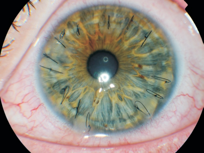
Flap Striae Columbia Ophthalmology

A Guide To Lasik And Refractive Surgery Options Premier Eye Associates
Laser Lasik Eye Surgery Reviews Lasik Eye Surgery Flap Healing
Blog Page 2 Of 11 Nvision Eye Centers

Management Of A Traumatic Flap Dislocation Seven Years After Lasik

Slit Lamp Photograph Of The Injured Eye Five Days After Flap Download Scientific Diagram
.jpg)
Lasik Flap Dislocation The Flap Never Heals

Services Mahajan Eye Centre
/flap-detach-bullous-keratopathy.jpg)
Lasik Flap Dislocation The Flap Never Heals

Full Text Traumatic Corneal Flap Displacement After Laser In Situ Keratomileusis Imcrj

Circumferential Epithelial Defect At Flap Margins In Patient With Adenoviral Conjunctivitis And Previous Lasik Eye

Eyeworld Management Of Rare Post Lasik Complications

Pdf Traumatic Corneal Flap Dislocation Four Years After Lasik Semantic Scholar
.jpg)
Lasik Flap Dislocation The Flap Never Heals
Early Adopter Chronic Sufferer

Smile Vs Lasik

Troubleshooting In Lasik Eye News
/detached-lasik-flap-2.jpg)
Lasik Flap Dislocation The Flap Never Heals

Pdf Traumatic Corneal Flap Dislocation Four Years After Lasik Semantic Scholar
Late Traumatic Flap Dislocation Seven Years After Femtosecond Laser Assisted In Situ Keratomileusis
Effects Of The Lasik Flap Thickness On Corneal Biomechanical Behavior A Finite Element Analysis Bmc Ophthalmology Full Text
Smile Eye Surgery Costs Safety Recovery Timelines
Lasik Jonesblog

Smile Five Years On Does It Really Have A Role Mivision
Navigating The Lasik Postoperative Journey Collaborativeeye
Treating Epithelial Ingrowth After Lasik

The Resurgence Of Post Lasik Epithelial Ingrowth William Nc Nixon Hk Pudukadan D Bhat L Kerala J Ophthalmol
Late Onset Dehiscence Of Lasik Flap Poster Number P 190 Authors Hyunjin Jane Kim Cary M Silverman Category Keratorefractive Authors Have No Financial Ppt Download

Eyeworld Focusing On Lasik Femtosecond Side Cuts

Lasik Vs Prk What The Lasik Doctor Doesn T Tell You Is This Your Homework

Troubleshooting In Lasik Eye News

Lasik Surgery In Delhi Laser Eye Treatment Hospital Doctor Cost

Ophthalmology Management Early Intervention For Post Lasik Trouble

Treatment Of Traumatic Lasik Flap Dislocation And Epithelial Ingrowth With Fibrin Glue American Journal Of Ophthalmology

Top 10 Reasons Why You Should Not Get Lasik Eye Surgery Omg Top Tens List

Laser In Situ Keratomileusis In 12 A Review Sutton 14 Clinical And Experimental Optometry Wiley Online Library

Therapeutic Flap Amputation For Atypical Lasik Flap And Interface Abnormalities

Late Traumatic Lasik Flap Loss During Contact Sport Sciencedirect

Epithelial Ingrowth Following Laser In Situ Keratomileusis Lasik Prevalence Risk Factors Management And Visual Outcomes Bmj Open Ophthalmology

Ophthalmology Management Refractive Surgery With A Smile
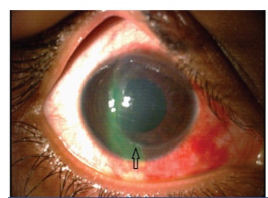
Jcdr Closed Globe Injury Displaced Flap Partial Thickness Laceration Of Cornea
.jpg)
Lasik Flap Dislocation The Flap Never Heals

Lasik Complications And Their Management Ento Key
Lvc No Longer Out Of Bounds For Athletes

Reasons For Lasik Enhancement Washington Dc
/flap_edge_dye(425).jpg)
Lasik Flap Dislocation The Flap Never Heals

Characterisation Of Corneal Fibrotic Wound Repair At The Lasik Flap Margin British Journal Of Ophthalmology

A Clear Look At The Lasik Flap

Which Is Better Lasik Or Prk Schulze Vision Surgery Center

Recovery Time After Laser Eye Surgery What To Expect Dougherty Laser

Pdf Management Of A Traumatic Flap Dislocation Seven Years After Lasik

Full Text Traumatic Corneal Flap Displacement After Laser In Situ Keratomileusis Imcrj

Z Ruo9rcshewlm

Late Traumatic Lasik Flap Loss During Contact Sport Sciencedirect

Corneal Flap Complications In Refractive Surgery Part 1 Development Of An Experimental Animal Model Sciencedirect

Epithelial Ingrowth After Late Traumatic Femtosecond Laser Assisted Laser In Situ Keratomileusis Flap Dislocation Sciencedirect
/ectasia_flap_margin(425).jpg)
Lasik Flap Dislocation The Flap Never Heals
/detached-lasik-flap(640).jpg)
Lasik Flap Dislocation The Flap Never Heals

Late Post Traumatic Flap Dislocation And Macrostriae After Laser In Situ Keratomileusis Sinha R Shekhar H Tinwala S Gangar A Titiyal Js Oman J Ophthalmol
Late Traumatic Flap Dislocation Seven Years After Femtosecond Laser Assisted In Situ Keratomileusis

Small Incision Lenticule Extraction Smile It S What S New In Laser Vision Correction Harvard Health Blog Harvard Health Publishing

Prk Vs Lasik Eye Surgery

Traumatic Flap Dislocation 10 Years After Lasik Case Report And Literature Review Sciencedirect

Something To Smile About

Femtosecond Laser Eases Lasik Fears

Lasik Interface Primary Complications

Figure 4 From Satisfactory Clinical Outcome Following Delayed Repositioning Of A Traumatic Post Lasik Flap With Dislocation And Shrinkage Managed By Irrigation Stretching And Debridement Semantic Scholar
Laser Eye Surgery Primer Lasik Vs Prk Is This Your Homework

Traumatic Corneal Flap Displacement Five Years After Laser In Situ Keratomileusis Semantic Scholar
Intraoperative Flap Complications In Lasik Prevention And Management Of Free Flaps Springerlink

Hd Corneal Flap Displacement From Eye Trauma After Lasik Em Rap

Lasik Complications And Their Management Ento Key

Eyeworld Management Of Rare Post Lasik Complications

Late Post Traumatic Flap Dislocation And Macrostriae After Laser In Situ Keratomileusis Sinha R Shekhar H Tinwala S Gangar A Titiyal Js Oman J Ophthalmol

Lasik And Flap Complications Salt Lake City Ut Risks

Corneal Flap Dehiscence After Screwdriver Trauma Nejm


